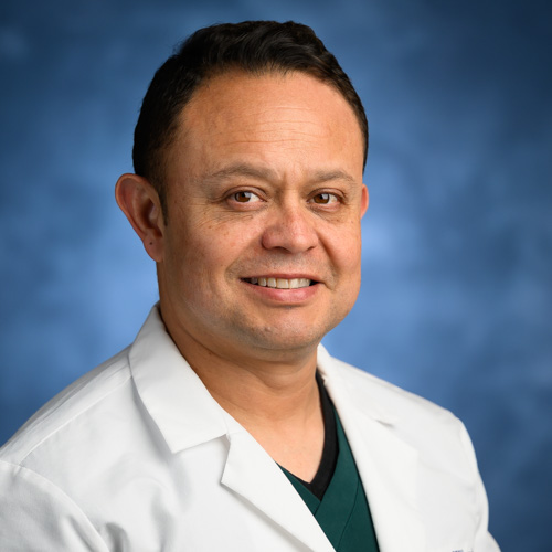
Lenin Villamizar Martinez DVM, Dipl. AVDC

Bio
Dr. Lenin Arturo Villamizar-Martinez is an Associate Professor, Resident director, and head of the Dentistry and Oral Surgery Service, at North Carolina State University College of Veterinary Medicine. Diplomate of the American Veterinary Dental College since 2019. He completed his veterinary degree at De La Salle University, Bogotá, Colombia, in 2003. After graduation, he spent three years in companion animal practice in Bogotá before moving in 2006 to Brazil to perform his master’s, Ph.D., and postdoctoral studies at the Veterinary and Animal Science School of the University of São Paulo. During this time, he also participated as a research fellow and clinical instructor collaborator in the Diagnostic Imaging Service and the Comparative Dentistry laboratory of the School of Veterinary Medicine and Animal Science of the University of Sao Paulo. Dr. Villamizar moved to Philadelphia in 2014 to perform his Dentistry and oral surgery residency at the University of Pennsylvania School of Veterinary Medicine. He is the recipient of the Journal of Veterinary Dentistry Editor’s Award 2016 for the most outstanding manuscript in the Journal of Veterinary Dentistry. His clinical and research interests include maxillofacial trauma, oncology surgery, and diagnostic imaging of the dental and maxillofacial structures and the temporomandibular joint.
Affiliations
Diplomate of the American Veterinary Dental College – AVDC
Education
Postdoctoral Fellow University of São Paulo (Brazil) 2014
PhD University of São Paulo (Brazil) 2012
MS University of São Paulo (Brazil) 2008
DVM De La Salle University (Bogotá-Colombia) 2003
Area(s) of Expertise
Maxillofacial trauma, oncology surgery, and diagnostic imaging of the dental and maxillofacial structures and the temporomandibular joint.
Publications
- Canine and feline dental disease , Thall's Textbook of Veterinary Diagnostic Radiology (2025)
- Halitosis and ptyalism , Ettinger's Textbook of Veterinary Internal Medicine (2024)
- Utilization of an acrylic-based obturator for managing an oronasal communication in a ferret , 38th Veterinary Dental Forum, 2024, Palm Springs-CA. Proceedings (2024)
- Assessment of the Occupational Radiation Dose from a Handheld Portable X-ray Unit During Full-mouth Intraoral Dental Radiographs in the Dog and the Cat – A Pilot Study , Journal of Veterinary Dentistry (2023)
- Evaluation of 3D-Printed Dog Teeth for Pre-clinical Training of Endodontic Therapy in Veterinary Dentistry , Journal of Veterinary Dentistry (2023)
- Caudal and middle segmental mandibulectomies for the treatment of unilateral temporomandibular joint ankylosis in cats , Journal of Feline Medicine and Surgery Open Reports (2022)
- Computed Tomography Assessment of Tidal Lung Overinflation in Domestic Cats Undergoing Pressure-Controlled Mechanical Ventilation During General Anesthesia , Frontiers in Veterinary Science (2022)
- Morphometry and Morphology of the Articular Surfaces of the Medial Region of the Temporomandibular Joint in the Felis Catus (Domestic cat)-A Cone Beam Computed Tomography Study , JOURNAL OF VETERINARY DENTISTRY (2022)
- Diagnostic Imaging of Oral and Maxillofacial Anatomy and Pathology , Veterinary Clinics of North America: Small Animal Practice (2021)
- Localization of the First Mandibular Molar Roots in Relationship to the Mandibular Canal in Small Breed Dogs—A Tomography Imaging Study , Frontiers in Veterinary Science (2021)
Grants
Dr. Martinez will be responsible for all aspects of the canine imaging study at the performance site as described in the attached NC State IACUC Protocol #20-133, approved 16-Mar -2020 Specifically, Dr. Martinez will be in charge of: - Identifying and enrolling 25 patient animals (dogs) to participate - Injecting the contrast and performing the imaging per the protocol referenced above. o Approximately 10 animals to be imaged post-injection using a varying time between injection and scan using the 20s imaging technique on VetCAT. o Approximately 15 animals to be imaged concurrent with injection while varying scan times and injection times. - Evaluation of images of the enhanced vessels and tissues, tumor visibility (if applicable), vessel continuity, degree of enhancement, distinction between arterial and venous anatomy, presence or absence of artifact, and clinical utility. Comparison to conventionally acquired CT images if available. - Prepare a brief summary report of relevant observations - Ultimately responsible for all patient care for all enrolled subjects. The purpose of this project is to establish cranial contrast CT imaging protocols in an animal (canine) organism providing a level of realism beyond what is achievable in a laboratory phantom model.
Groups
Honors and Awards
- Journal of Veterinary Dentistry Editor’s Award 2016 for the most outstanding manuscript in the Journal of Veterinary Dentistry